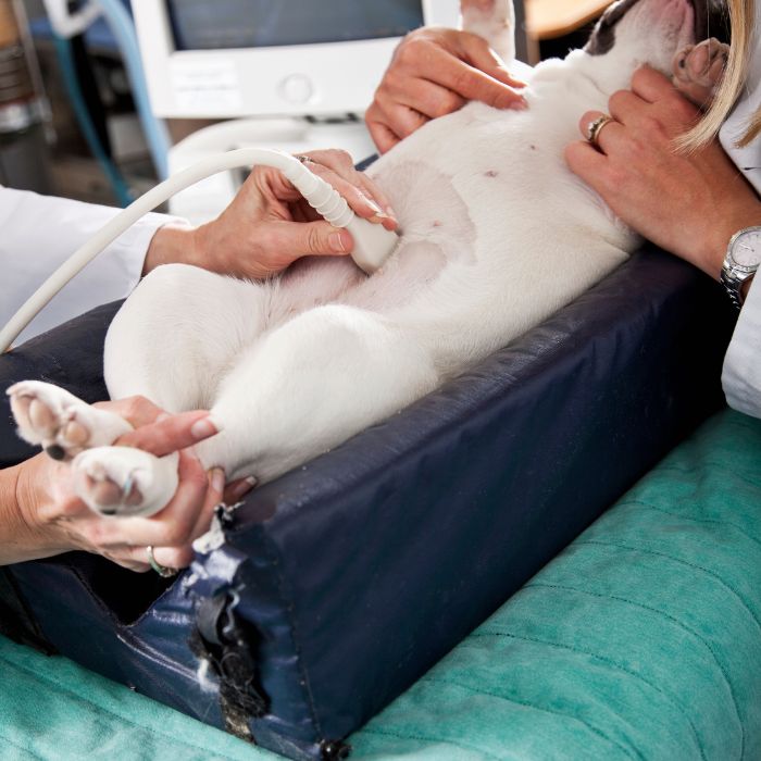
Pet Ultrasound
The internal organs of an animal can be examined using veterinary ultrasonography.

Pet Ultrasound
Ultrasound helps us to explore soft tissue illnesses that may not show up well on X-rays in a painless and non-invasive manner. It is a valuable tool for diagnosing a wide range of illnesses, including heart disease, liver disease, kidney disease, and cancer. It can also be used to identify pregnancy and track the development of fetuses. Ultrasounds are performed in-house and in cooperation with a board-certified internist.
Pet ultrasound, also known as ultrasonography, is a powerful diagnostic tool that has revolutionized veterinary medicine. By using sound waves, veterinarians can obtain real-time images of your pet’s internal organs, providing invaluable insights into their health.
This non-invasive procedure allows our veterinarians to examine organs such as the heart, liver, kidneys, bladder, and reproductive organs. It is particularly effective in identifying abnormalities like tumors, cysts, stones, or inflammation and aiding in diagnosing conditions such as urinary tract infections, gastrointestinal disorders, and heart diseases.
The real-time imaging capabilities of pet ultrasound allow our veterinarians to assess organ functionality, guide biopsies, assist in procedures, and monitor treatment effectiveness. Unlike other diagnostic methods, pet ultrasound is generally well-tolerated by pets and does not require anesthesia in most cases, reducing risks and promoting faster recovery.
In summary, pet ultrasound is an invaluable tool in veterinary care. Its ability to provide detailed, real-time images of your pet’s internal organs enables veterinarians to make accurate diagnoses and develop targeted treatment plans, ultimately ensuring the best possible health outcomes for your furry companions.
